Pes Anserine Bursitis
1. Introduction
The pes anserinus (“goose’s foot”) is the combined tendinous insertion of the sartorius, gracilis and semitendinosus tendons at their attachment to the tibia (shin bone). The primary action of these muscles is to flex the knee and to resist the knee falling inwards (valgus strain) (1,2). The risk of pes anserine bursitis increases with excessive valgus stress. This can occur due to ligamentous instability, meniscal injuries, muscular imbalances or knock knee deformity (4).
A bursa is a cavity-like structure lined with synovial tissue that cushions and assists the motion of tissues that cross it during joint movement. The pes anserinus bursa lies between the pes anserine insertion (“goose’s foot”) and the periosteum of the shin bone and may become inflamed due to overuse. It is relatively uncommon but may occur in swimmers, runners and cyclists (2).
Pes anserine pain syndrome is a more generic term that is used to refer to medial (inside) knee pain, which may or may not include inflammation of the bursa.
Frequently Asked Questions
- Pes anserine bursitis is an inflammation of the bursa (fluid filled sac) located between the shinbone (tibia) and three muscle tendons that attach at a shared site on the inside of the knee.
- Uncommon in the general population.
- There is an established link with knee osteoarthritis and other knee joint conditions (1).
- More likely in the sporting population (2).
- No
- This condition typically occurs secondary to other knee injury and therefore it usually improves with resolution of the primary knee condition.
- If this condition occurs in isolation, it will normally resolve with physiotherapy input and self-management (3).
- More common in overweight middle-aged women (2).
- Particularly those involved in sports such as running, basketball and racquet sports (2).
- Pain and tenderness to the touch.
- Swelling and warmth.
- Tightness of the soft tissue attachments of the knee.
- Reduced range of movement due to pain and tightness.
- In the early stage of management good practice now involves use of the Peace & Love protocol see below for more explanation.
- Maintain weight-bearing as tolerated.
- Physiotherapy guided management is very beneficial in the rehabilitation of this condition (3).
- Typically, rehabilitation of this injury will take between 6-8 weeks (3).
- Recovery timeframes and limitations will be influenced by other pre-existing knee conditions.
We recommend consulting a musculoskeletal physiotherapist to ensure exercises are best suited to your recovery. If you are carrying out an exercise regime without consulting a healthcare professional, you do so at your own risk. Book online with us today to get a programme tailored to your specific needs.
2. Signs and Symptoms
Symptoms are typically localised to the inside of the knee (2). These can include:
- Pain and tenderness to the touch.
- Swelling and warmth.
- Tightness of the soft tissue attachments of the knee.
- Reduced range of movement due to pain and tightness.
3. Causes
This condition typically occurs because of overuse – when the relevant muscles and tendons are repetitively loaded in flexed positions such as running. This increases the level of friction and pressure on the underlying bursa triggering an inflammatory response. This can present as pain, swelling and weakness in patients. Pes anserine bursitis can also occur due to direct trauma or biomechanical abnormalities such as knock knees (4).
Other causes include underlying knee pathology such as osteoarthritis or medial collateral ligament injury. A sudden change in physical activity levels can also contribute to the onset of this condition (1,2).
4. Risk Factors
This is not an exhaustive list. These factors could increase the likelihood of someone developing a pes anserine bursitis (1,2). It does not mean everyone with these risk factors will develop symptoms.
- Overuse injury
- Valgus knee deformity
- Muscle weakness
- Underlying arthritic changes
- Direct trauma
- Obesity
- Diabetes
5. Prevalence
A study into knee pain in 500+ patients found that pes anserine bursitis was detected on MRI in 2.5% of patients. Research indicates that less than 1% of the general population will present with pes anserine bursitis (5).
Other research suggests pes anserinus pain prevalence is rare in the general population but is most common when associated with osteoarthritis in the same knee (1).
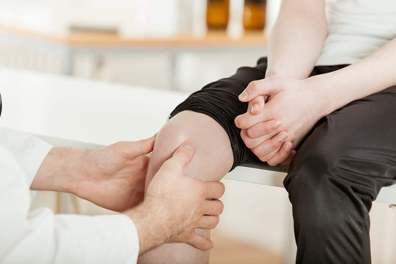
6. Assessment & Diagnosis
A musculoskeletal physiotherapist can provide you with an accurate and timely diagnosis by obtaining a detailed history of your symptoms. A series of physical tests might be performed as part of your assessment to exclude other potentially involved structures and gain a greater understanding of your physical abilities to help facilitate an accurate working diagnosis.
Imaging is not usually required to make a pes anserinus bursitis diagnosis, but your physiotherapist may request further imaging if significant intra-articular pathology needs to be excluded; predominately arthritis.
7. Self-Management
As part of your treatment, your physiotherapist will help you understand the condition and what needs to be implemented to effectively manage pes anserine bursitis. This will include activity modification strategies as well as other useful treatments aimed at reducing discomfort. Regular adherence to a condition specific rehabilitation programme is important in the management of this condition. It should be noted that rehabilitation exercises are not always a quick fix but if adhered to on a consistent basis (weeks to months), over time they have been shown to yield positive outcomes.
Initial management will focus on reducing pain and limiting swelling. You may be advised to rest and use ice regularly in the initial stages to help manage pain and swelling.
Weight loss and muscle strengthening exercises are important in supporting recovery and helping with the long-term resolution of symptoms (2, 3, 4).
8. Rehabilitation
Your physiotherapist will advise on a specific exercise programme which is tailored to address any muscular weakness or imbalances, as well as ensuring the knee joint has an optimal range of movement. A more general approach to lifestyle changes may also be addressed during your consultation, including weight management advice to compliment optimal knee function.
Further physiotherapy treatment modalities may be used to assist with pain relief when pain persists (2, 3, 5).
Guidance and pacing on return to sport is imperative due to the overload nature of this injury. Your physiotherapist will discuss this with you prior to the end of your rehabilitation.
9. Pes Anserine Bursitis
Rehabilitation Plans
Our team of expert musculoskeletal physiotherapist have created rehabilitation plans to enable people to manage their condition. If you have any questions or concerns about a condition, we recommend you book an consultation with one of our clinicians.
What Is the Pain Scale?
The pain scale or what some physios would call the Visual Analogue Scale (VAS), is a scale that is used to try and understand the level of pain that someone is in. The scale is intended as something that you would rate yourself on a scale of 0-10 with 0 = no pain, 10 = worst pain imaginable. You can learn more about what is pain and the pain scale here.
This programme focuses on ensuring you achieve optimal range of movement and good muscle strength. We suggest you perform this once a day for approximately 1-3 weeks as your symptoms allow. The exercises are unlikely to be painful but if there is any discomfort, aim to keep this below a 3 on the scale below.
- 0
- 1
- 2
- 3
- 4
- 5
- 6
- 7
- 8
- 910
- 0
- 1
- 2
- 3
- 4
- 5
- 6
- 7
- 8
- 910
These exercises are important in re-establishing the good strength of the major muscle groups in the lower limb, particularly the quadriceps. We suggest you perform this once a day for approximately 4 – 8 weeks as your symptoms allow. These exercises should be performed with minimal pain (3/10) but you may experience some tightness on the inside of your knee if swelling remains.
- 0
- 1
- 2
- 3
- 4
- 5
- 6
- 7
- 8
- 910
10. Return to Sport / Normal life
For patients targeting a high level of function or return to sport we would encourage a consultation with a physiotherapist as you will likely require further progression beyond the advanced rehabilitation stage. Before returning to sport, a rehabilitation programme should incorporate sport-specific exercises. This might include things like bounding, cutting, and sprinting exercises.
As part of a multi-modal treatment approach, your Physiotherapist may also use a variety of other pain-relieving treatments to support symptom relief and recovery. During rehabilitation, you may benefit from further assessment to ensure progression and appropriate tailoring of treatment. Ongoing support and advice will allow you to self-manage and prevent future re-occurrence.
11. Other Treatment Options
An onward referral to orthopaedics may be required should significant intra-articular pathology be suspected such as an ACL injury or complex meniscal tear.
Steroid injections may be used in cases with severe nocturnal pain that have not responded to self-management (5).
12. Links for Further Reading
25 locations and counting across the UK
References
- Sapp GH, Herman DC. Pay Attention to the Pes Anserine in Knee Osteoarthritis. Curr Sports Med Rep. 2018 Feb;17(2):41.
- Bisciotti, Gian Nicola & Volpi, Piero. (2016). The Lower Limb Tendinopathies: Etiology, Biology and Treatment. 10.1007/978-3-319-33234-5.
- Sarifakioglu B, Afsar SI, Yalbuzdag SA, Ustaömer K, Bayramoğlu M. Comparison of the efficacy of physical therapy and corticosteroid injection in the treatment of pes anserine tendino-bursitis. J Phys Ther Sci. 2016 Jul;28(7):1993-7. [4]
- Mohseni M, Graham C. Pes Anserine Bursitis. [Updated 2020 Nov 26]. In: StatPearls [Internet]. Treasure Island (FL): StatPearls Publishing; 2020 Jan-. Available from: https://www.ncbi.nlm.nih.gov/books/NBK532941/
- Yoon HS, Kim SE, Suh YR, Seo YI, Kim HA. Correlation between ultrasonographic findings and the response to corticosteroid injection in pes anserinus tendinobursitis syndrome in knee osteoarthritis patients. J Korean Med Sci. 2005 Feb;20(1):109-12.[5]
Other Conditions in
Knees
Patellofemoral Pain Syndrome (PFPS)
Knee pain around the kneecap usually worse in static positions, squatting or kneeling.
Patellar Tendinopathy
Knee pain at the lower border of the kneecap which is also known as ‘jumper’s knee’.
Patella Dislocation
Patella dislocation is a knee injury in which the patella (kneecap) slips out of its normal position. The most common direction for the kneecap to dislocate is laterally or the outside. This is commonly associated with pain and swelling in the soft tissue tissues which may have been stretched or damaged. Patella subluxation refers to when the kneecap is only partially displaced and then returns to it’s normal location.
Osgood-Schlatter Disease
Pain in an area just below the knee on the shin bone, often with a lump.
Meniscus Injury
Structural knee injury, triggered either by a tear or through wear and tear.
Medial Collateral Ligament Sprain
The medial collateral ligament is on the inner side of the knee. It provides stability to the joint by preventing excessive side–to–side movement. It is possible to injure this ligament when a person is bearing weight, and the knee is forced inwards.
Lateral Collateral Ligament (LCL) Injury
The lateral collateral ligament is a strong ligament on the outside of the knee. A tear will only occur during a high force impact or twisting motion.
Knee Replacement Surgery
Replacement of the knee hinge joint, typically as a result of severe osteoarthritis or trauma.
Knee Osteoarthritis
Common age related changes to the structure of the knee joint which may be associated with pain, stiffness and loss of function.
Iliotibial Band Syndrome
Presents as pain on the outside of the knee, normally occurring because of overload due to prolonged or repeated bouts of exercise.
Hamstring Strain/Tear
An over-stretch or tear to one or more of the muscles located at the back of the thigh.
Femoral Nerve Radiculopathy
This is where the nerve that supplies the front of the leg is irritated and causes pain/numbess.
Fat Pad Impingement
A rare condition affecting the adipose (fat) tissue that sits under the kneecap (patella) between the joint spaces of the knee.
Degenerative Meniscus
Seen to be normal as we age, but in some situations can result in knee aches, pain or joint swelling.
Bowed Knees
A condition in which the legs are bowed outwards leaving a greater space in between your knees.
Benign Joint Hypermobility Syndrome
Common age related changes to the structure of the knee joint which may be associated with pain, stiffness and loss of function.
Baker’s Cyst
Swelling in the popliteal space (space behind the knee) that causes a visible lump.
Anterior Cruciate Ligament (ACL) Injury
Injury to a major stability ligmant in the knee, normally occuring following a significant twisting injury.