Fat Pad Impingement
1. Introduction
Articular (relating to a joint) fat pads are accumulations of fatty soft tissue that occupy space and irregularities within a joint. They act as flexible protective cushions that accommodate the changing shape of the joint during movement (3).
The infrapatellar fat pad, also known as the Hoffa’s fat pad, sits behind and just below the kneecap between the shin (tibia) and thigh (femur) bones. The location is thought to expose the fat pad, making it more suspectable to injury, especially during the end of range knee extension (straightening) (1).
If pain is present at the front of the knee, around the bottom of the kneecap and is worse when the knee is fully extended, the infrapatellar fat pad might be the source of discomfort.
The movement of knee extension (in symptomatic cases) causes the fat pad to become impinged, hence the reason why some clinicians will refer to this condition as infrapatellar (beneath the kneecap) fat pad impingement syndrome. The fat pad also has a rich supply of nerves which contribute to the discomfort levels often experienced with this condition (2).
Frequently Asked Questions
- Fat pad impingement, otherwise known as Hoffa’s syndrome, is a rare condition affecting the adipose (fat) tissue that sits under the kneecap (patella) between the joint spaces of the knee.
- In the general population, it affects less than 1% of people who present with anterior (front) knee pain (4), which makes it a very rare condition.
- No.
- With the right rehabilitation approach fat pad impingement generally recovers well.
- This condition is not linked to other serious pathology.
- It is more common in women than in men (1).
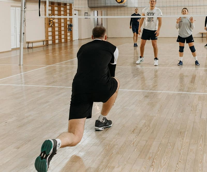
- Those that take part in jumping sports such as basketball and volleyball, etc.
- Individuals with increased flexibility of their ligaments and joints (2,4), e.g. hypermobility syndrome.
- Localised pain just below the kneecap with activity e.g. jumping, running, squatting and ascending or descending stairs (3).
- Increased pain when the knee is fully extended (straightened) as the fat pad can become pinched (3).
- Modify your activity – avoiding jumping sports and sustained end of range knee extension (leg full straightened) whilst the knee is painful.
- Progressive and appropriate loading of the muscles around the knee has been shown to be one of the most effective treatments.
- Advice from a qualified physiotherapist will be helpful in most cases.
- Initial recovery can take between 8-12 weeks and full recovery between 3-6 months (6, 7).
- If left untreated, symptoms can return if you go back to usual activities, without having gone through an appropriate rehabilitation programme (6).
We recommend consulting a musculoskeletal physiotherapist to ensure exercises are best suited to your recovery. If you are carrying out an exercise regime without consulting a healthcare professional, you do so at your own risk. Book online with us today to get a programme tailored to your specific needs.
2. Signs and Symptoms
- Pain is usually described as being felt behind or just below the knee cap.
- Placing increased pressure through the knee may cause a worsening of symptoms such as when ascending and descending stairs, jumping and running.
- In more severe and irritable cases, patients can also present with a degree of localised swelling and movement can be restricted (3).
3. Causes
Most commonly this condition is thought to develop gradually over time following repetitive overextension of the knee. Some individuals may be more susceptible to developing this injury if they have perpetuity towards knee hyperextensions, such as those who partake in jumping sports and those with ligament and joint laxity conditions such as hypermobility syndrome. In other instances, this condition can start following a direct blow to the knee (1, 3).
4. Risk Factors
This is not an exhaustive list. These factors could increase the likelihood of someone developing fat pad impingement. It does not mean everyone with these risk factors will develop symptoms.
- Predominantly seen in young women (1).
- Those that take part in jumping sports.
- Ligamentous and joint laxity conditions such as hypermobility syndrome – fat pad more at risk of impingement due to increased risk of knee hyperextension (1).
5. Prevalence
Prevalence is not fully understood however, studies reported isolated fat pad injuries in 1% of people suffering from anterior knee pain (1), which makes it a very rare condition.
6. Assessment & Diagnosis
Musculoskeletal physiotherapists and other appropriately qualified healthcare professionals can provide you with a diagnosis by obtaining a detailed history of your symptoms. A series of physical tests might be performed as part of your assessment to rule out other potentially involved structures and gain a greater understanding of your physical abilities to help facilitate an accurate working diagnosis.
Your treating clinician will want to know how your condition affects you day-to-day so that treatment can be tailored to your needs and personalised goals can be established. Intermittent reassessment will ascertain if you are making progress towards your goals and will allow appropriate adjustments to your treatment to be made. Imaging studies like MRIs or ultrasound scans are usually not required to achieve a working diagnosis, but in unusual presentations, they may be warranted.
7. Self-Management
Your treating clinician will help you understand your condition so that you are able to help support your recovery. Initial treatment should focus on reducing pain and inflammation. Dependent on the severity and physical limitations, it is recommended that you limit or avoid aggravating tasks. Some examples include reducing prolonged periods of standing, remaining vigilant for not over-extending the knee and avoiding sports that exacerbate symptoms. If your symptoms do not improve to the point where you feel you are able to self-manage then it would be encouraged to seek further input from an appropriately qualified musculoskeletal specialist. We encourage the utilisation of methods highlighted in this PEACE & LOVE acronym (see below) which can be implemented for early recovery but should always be guided by an appropriately qualified musculoskeletal specialist (8).
Protect: the injury the first few days, avoid activities that increase pain.
Elevate: the leg higher than the heart as often as possible.
Avoid anti-inflammatories: they may reduce tissue healing in the early stage.
Compression: can help reduce swelling.
Education: your body knows best, avoid unnecessary treatments.
&
Load: let pain guide your return to normal activities.
Optimism: remain confident and positive.
Vascularisation: choose a pain-free exercise that elevates your heart rate.
Exercise: restore strength by adopting an active approach to recovery.
8. Rehabilitation
Your clinician may use certain taping techniques on the symptomatic knee which may ease symptoms. This will often be combined with a condition-specific strengthening programme. Exercises will be individualised to your needs and should be progressed as you recover. Regular reassessment will allow modifications to be made and will ensure you are making progress towards your goals.
Below are three rehabilitation programmes created by our specialist musculoskeletal physiotherapists. In some instances, a one-to-one assessment is appropriate to individually tailor targeted rehabilitation. However, these programmes provide an excellent starting point as well as clearly highlighting exercise progression.
9. Fat Pad Impingement
Rehabilitation Plans
Our team of expert musculoskeletal physiotherapist have created rehabilitation plans to enable people to manage their condition. If you have any questions or concerns about a condition, we recommend you book an consultation with one of our clinicians.
What Is the Pain Scale?
The pain scale or what some physios would call the Visual Analogue Scale (VAS), is a scale that is used to try and understand the level of pain that someone is in. The scale is intended as something that you would rate yourself on a scale of 0-10 with 0 = no pain, 10 = worst pain imaginable. You can learn more about what is pain and the pain scale here.
In this early plan, our focus is on maintaining good movement through the area whilst we try and reduce the severity of the symptoms. Pain should not exceed 3/10 on your perceived pain scale whilst completing this exercise programme.
- 0
- 1
- 2
- 3
- 4
- 5
- 6
- 7
- 8
- 910
This is the next progression. Here we start to try and regain strength in the thigh muscles to ensure optimal strength and stability around the knee. Pain should not exceed 4/10 whilst completing this exercise programme.
- 0
- 1
- 2
- 3
- 4
- 5
- 6
- 7
- 8
- 910
This programme is a further progression increased emphasis on the strength of the area and moving towards more functional and dynamic tasks.
Pain should not exceed 4/10 whilst completing this exercise programme.
- 0
- 1
- 2
- 3
- 4
- 5
- 6
- 7
- 8
- 910
10. Return to Sport / Normal life
For patients wanting to achieve a higher level of function or return to sport, we would encourage a consultation with a musculoskeletal physiotherapist as you will likely require further progression beyond the advanced rehabilitation stage. Before returning to the sport, a rehabilitation programme should incorporate plyometric based exercises which might include things like bounding, quick changes in direction of movement and sprinting exercises.
11. Other Treatment Options
Corticosteroid injection may provide temporary pain relief, but this should only be considered as a last resort if appropriate and progressive conservative management has failed. Even if conservative management does not achieve a 100% improvement, careful consideration is heavily encouraged as in some cases injections cause more harm than good. Surgery may be recommended but this should be the last option if all other treatment attempts have been exhausted (5).
25 locations and counting across the UK
References
- Larbi, A., Cyteval, C., Hamoui, M., Dallaudiere, B., Zarqane, H., Viala, P. and Ruyer, A. (2014). Hoffa’s disease: A report on 5 cases. Diagnostic and interventional imaging, 95(11), 1079-1084.
- Hannon, J., Bardenett, S., Singleton, S. and Garrison, J.C. (2016). Evaluation, treatment, and rehabilitation implications of the infrapatellar fat pad. Sports health, 8(2), 167-171.
- Jarraya, M., Diaz, L.E., Roemer, F.W., Arndt, W.F., Goud, A.R. and Guermazi, A. (2018). MRI findings consistent with peripatellar fat pad impingement: how much related to patellofemoral maltracking?. Magnetic Resonance in Medical Sciences, 17(3), 195.
- Kumar, D., Alvand, A. and Beacon, J.P. (2007). Impingement of infrapatellar fat pad (Hoffa’s disease): results of high-portal arthroscopic resection. Arthroscopy: The Journal of Arthroscopic & Related Surgery, 23(11), 1180-1186.
- Morgans, E. (2020). Infrapatellar fat pad syndrome. BUPA. Date viewed [18.01.2021]. Available from <https://www.bupa.co.uk/health-information/knee-clinic/knee-conditions/infrapatellar-fat-pad-syndrome>
- Borja, M.J., Jose, J., Vecchione, D., Clifford, P.D. and Lesniak, B.P. (2013). Prefemoral fat pad impingement syndrome: identification and diagnosis. The American Journal of Orthopedics, 42(1), 9-11.
- Dragoo, J.L., Johnson, C. and McConnell, J. (2012). Evaluation and treatment of disorders of the infrapatellar fat pad. Sports medicine, 42(1), 51-67.
- Dubois, B. & Esculier, J. (2020); “Soft-tissue injuries simply need PEACE and LOVE”, British journal of sports medicine, 54(2), 72-73.
Other Conditions in
Knees, Orthopaedics
Patellofemoral Pain Syndrome (PFPS)
Knee pain around the kneecap usually worse in static positions, squatting or kneeling.
Patellar Tendinopathy
Knee pain at the lower border of the kneecap which is also known as ‘jumper’s knee’.
Osgood-Schlatter Disease
Pain in an area just below the knee on the shin bone, often with a lump.
Meniscus Injury
Structural knee injury, triggered either by a tear or through wear and tear.
Medial Collateral Ligament Sprain
Lateral Collateral Ligament (LCL) Injury
The lateral collateral ligament is a strong ligament on the outside of the knee. A tear will only occur during a high force impact or twisting motion.
Knee Replacement Surgery
Replacement of the knee hinge joint, typically as a result of severe osteoarthritis or trauma.
Knee Osteoarthritis
Common age related changes to the structure of the knee joint which may be associated with pain, stiffness and loss of function.
Iliotibial Band Syndrome
Presents as pain on the outside of the knee, normally occurring because of overload due to prolonged or repeated bouts of exercise.
Hamstring Strain/Tear
An over-stretch or tear to one or more of the muscles located at the back of the thigh.
Femoral Nerve Radiculopathy
This is where the nerve that supplies the front of the leg is irritated and causes pain/numbess.
Degenerative Meniscus
Seen to be normal as we age, but in some situations can result in knee aches, pain or joint swelling.
Bowed Knees
A condition in which the legs are bowed outwards leaving a greater space in between your knees.
Benign Joint Hypermobility Syndrome
Common age related changes to the structure of the knee joint which may be associated with pain, stiffness and loss of function.
Baker’s Cyst
Swelling in the popliteal space (space behind the knee) that causes a visible lump.
Anterior Cruciate Ligament (ACL) Injury
Injury to a major stability ligmant in the knee, normally occuring following a significant twisting injury.