Patella Dislocation
1. Introduction
The first episode of patella dislocation occurs in patients younger than 20 years in about 70% of cases (10). Patella dislocations can fall into two categories, traumatic and non-traumatic. A traumatic lateral patella dislocation occurs when twisting on a planted foot or if enough force is generated on the patella to disturb the MPFL (medial patella-femoral ligament). The forces must also be enough that the quadriceps and the depth of the trochlea (groove in which the patella sits) can no longer hold the patella in place. The patella may fully dislocate at this time or partially dislocate (subluxation) and pop back into place. Subluxations are more frequently mistaken with other injuries like an ACL tear. A thorough investigation including standard image X-ray is needed. Occasionally an MRI will be used to assess soft tissue structures, or a CT scan to look for smaller bone fragmentations.
Non-Traumatic dislocations or subluxations occur when the MPFL has previously been disturbed, genetic differences are found in the trochlea (thigh groove) and/or patella position; or due to changes to the patella on femoral (thigh bone) joint surface. Your physiotherapists understanding of the impact of cartilage or bony deformities, the history, age, height and weight allows them to make an informed choice on the appropriateness of conservative management or surgical intervention (4,9).
Frequently Asked Questions
- Commonly known as a dislocated kneecap. It occurs when the kneecap (patella) moves out of its normal position within the groove of the thigh bone, usually sliding to the outside of the knee.
- Lateral (outside of the knee) dislocations are accountable for around only 3% of traumatic knee injuries (10). This means it is not a common injury.
- A first-time (primary) patellar dislocation occurs in less than 0.05% people per year (4).
- Moderately, you should seek medical advice.
- If this is your first dislocation you are likely to make a full recovery with physiotherapy and rehabilitation (9).
- If you suffer from recurring dislocations your physiotherapist may liaise with a consultant to consider surgical options to stabilise the patella (9,10).
- Those aged between 10 and 17 years of age (9).
- Occurs in females more commonly than males (9).
- Those who have structural defects of the kneecap or the groove it sits in (8).
- Those who have suffered a previous patella dislocation (8).
- Impact sports, especially where the kneecap can be hit with force from the inside (1).
- Obvious deformity of the knee whereby the patella no longer sits central to the knee.
- A pop or snap when turning and accelerating with swelling soon following.
- Post injury pain, stiffness, swelling and a strange feeling to the movement of the kneecap, especially if it is unstable (7).
- With any major trauma you should seek emergency help or visit the local A&E department for them to put your patella back in place and get checked out for possible fractures.
- In the early stage of management good practice now involves use of the Peace & Love protocol (2).
- Complete an appropriate strengthening programme before returning to hobbies and sport (2).
- Range of motion is expected to recover in the first 6 weeks.
- Return to sport that includes change of direction and twisting may take more than 6 months (9).
- There are several factors involved in recovery including; age, gender, weight, damage to ligament structures, damage to patella or groove.
- Squatting and kneeling may be challenging long-term depending on the extent of the initial injury and your rehabilitation (3).
We recommend consulting a musculoskeletal physiotherapist to ensure exercises are best suited to your recovery. If you are carrying out an exercise regime without consulting a healthcare professional, you do so at your own risk. Book online with us today to get a programme tailored to your specific needs.
2. Signs and Symptoms
At times the injury will feel like a pop or snap when turning and accelerating. In an obvious dislocation the patella will not be in its central position on the knee. This will usually fall to the outside (lateral) part of the knee joint. This will need to be physically put back into place by a Physiotherapist, Paramedic or Doctor by creating medial forces on the patella whilst gradually extending the knee joint in a patellar reduction procedure. Following the injury, it is normal to experience pain, swelling and a strange feeling to move the kneecap, especially if it is unstable.
3. Causes
Traumatic dislocations or subluxations generally occur in two instances.
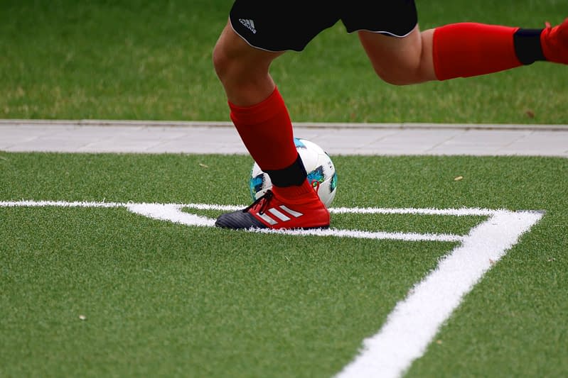
- Direct trauma to the inside of the knee forcing the patella laterally. This often disturbs the MPFL (medial patella-femoral ligament). The forces must also be enough that the medial fibres of the quadriceps, and the depth of the trochlea (thigh groove), can no longer hold the patella in place. An example of this is more common in football or rugby with a knee-high tackle.
- Twisting and accelerating on a planted foot. This may cause a lateral sheering force in the patella caused by the activation of the vastus lateralis quadricep muscle. This is more likely to cause a dislocation if you have already suffered with a past similar injury due to the disruption/ weakening of stabilisers of the joint. See below for risk factors on an unstable patellofemoral joint.
4. Risk Factors
This is not an exhaustive list. These factors could increase the likelihood of someone developing a patella dislocation injury. It does not mean everyone with these risk factors will develop symptoms.
- Those aged between 10 and 17 years of age (9).
- Occurs in females more commonly than males.
- Those who have genetic defects (8).
- Those who have suffered a previous patella dislocation (8).
- Impact sports especially where the kneecap can be hit from an inside (medial) force (1).
History of dislocations on one side were not found to be associated with an increased occurrence rate on the opposite side. In studies that reported on the presence of multiple risk factors, recurrence rates were 7.7% to 13.8% when no risk factors were present; increasing to 29.6% to 60.2% when 2 risk factors were present, and to 70.4% to 78.5% when 3 risk factors were present (8).
5. Prevalence
- Lateral (outside of the knee) dislocations are accountable for around only 3% of traumatic knee injuries (10). This means it is not a common injury.
- Other types of patella dislocations are described by the direction in which they occur superior (upward), inferior (downward) and medial (inside) but these are rare.
- A first-time (primary) patella dislocation occurs in less than 0.05% people per year (4).
6. Assessment & Diagnosis
A traumatic injury assessment should be received by your local emergency services. Following reduction of the patella you may be booked in for imaging or booked into an outpatient setting for further assessment by an Orthopaedic Consultant, Doctor or Physiotherapist. Assessment in an outpatient setting will involve a comprehensive assessment of yourself, hobbies, past injuries, mechanism of injury and possibly medical imaging. Tests to the knee by passively applying pressure to the patella to check for apprehension (how you feel) and signs of instability/ligament disruption may then be undertaken with your permission.
You may be diagnosed with an acute or recurrent dislocation or subluxation (partial dislocation) and may be classified further depending on your clinician’s experience. At this time early management strategies can be implemented and rehabilitation programmes guided on. Further referrals to orthopaedics and imaging may be considered in certain cases where exercise rehabilitation is not successful.
7. Self-Management
It is recommended to seek medical attention following dislocation to rule out damage to bony, ligamentous and cartilaginous structures that may require rest. Following the PEACE & LOVE sub-acute management strategies can be implemented for early recovery (2).
8. Rehabilitation
Rehabilitation will involve recovering range of motion in the first instance followed by loading quadricep attempting to bias vastus medialis (VMO/inside knee stabilisers) then eventually goal specific and return to play progressions for those involved in sports.
There is no evidence to suggest one rehabilitation exercise is more effective than another, or to suggest that open chain (non-weight bearing) or closed chain (weight bearing) exercises are preferred (9, 11). See below for possible rehabilitation ideas based on stages of your recovery developed by our team of physiotherapists.
9. Patella Dislocation
Rehabilitation Plans
Our team of expert musculoskeletal physiotherapist have created rehabilitation plans to enable people to manage their condition. If you have any questions or concerns about a condition, we recommend you book an consultation with one of our clinicians.
What Is the Pain Scale?
The pain scale or what some physios would call the Visual Analogue Scale (VAS), is a scale that is used to try and understand the level of pain that someone is in. The scale is intended as something that you would rate yourself on a scale of 0-10 with 0 = no pain, 10 = worst pain imaginable. You can learn more about what is pain and the pain scale here.
Early stages of rehabilitation should focus on swelling management, recovering range of motion and light core, glute and quad strengthening and proprioception work as able.
- 0
- 1
- 2
- 3
- 4
- 5
- 6
- 7
- 8
- 910
Mid stage rehabilitation should focus on weight bearing, alignment work, biasing VMO and general strengthening and proprioception exercise.
- 0
- 1
- 2
- 3
- 4
- 5
- 6
- 7
- 8
- 910
Advanced/return to sport stage should focus on sport related drills, change of direction work, plyometric and pivoting movements.
- 0
- 1
- 2
- 3
- 4
- 5
- 6
- 7
- 8
- 910
10. Return to Sport / Normal life
Return to sport can take anywhere from 6 months to 12 months. It is important not to rush back and risk recurrence of dislocation as this has shown to further de-stabilise the joint (8). Early introduction to sports-specific exercises can improve self-confidence, can increase compliance and facilitate a more rapid and safe return to the sport practice (10).
For patients wanting to achieve a high level of function or return to sport we would encourage a consultation with a physiotherapist as you will likely require further progression beyond the advanced rehabilitation stage. Before returning to sport, a rehabilitation programme should incorporate plyometric based exercises, this might include things like bounding, cutting, and sprinting exercises (10,11).
As part of a multi-modal treatment approach, your physiotherapist may also use a variety of other pain-relieving treatments to support symptom relief and recovery. Whilst recovering you might benefit from further assessment to ensure you are making progress and establish appropriate progression of treatment. Ongoing support and advice will allow you to self-manage and prevent future re-occurrence.
11. Other Treatment Options
Other treatment options include taping, insoles and knee sleeves though there is a lack of evidence to suggest how these strategies can help an unstable knee. These management tools may however help to increase awareness of the joint in space during rehabilitation and return to sport.
Taping may help with symptoms but has no effect on the alignment of the patella. Prolonged immobilisation has negative effects on muscles, bones, tendons and ligaments (10). Surgical management may be offered following first-time dislocation if there is obvious chondral (cartilage) damage on imaging or if conservative management has failed. This may include (9,10);
- Three-in-one procedure (lateral release, proximal vastus medialis realignment and plasty, and medial transfer of the medial one third of the patellar tendon).
- MPFL reconstruction.
- Tibial tubercle osteotomy.
- Trochleoplasty.
All surgical interventions involve varied levels of risk, recovery and outcomes depending on the person. Discuss thoroughly with your musculoskeletal physiotherapist and make an informed decision on any invasive treatments.
25 locations and counting across the UK
References
- Nomura E, Horiuchi Y, Kihara M. (2000). Medial patellofemoral ligament restraint in lateral patellar translation and reconstruction. Knee; 7:121–7
- Dubois B, Esculier J. A (2020) 54:72-73. The American Journal of Sports Medicine, 2020-08, Vol.48 (10), p.2552-2562 Soft-tissue injuries simply need PEACE and LOVE British Journal of Sports Medicine
- Dave M. Atkin, Donald C. Fithian, Kent S. Marangi, Mary Lou Stone, Barbara E. Dobson, and Cerrah Mendelsohni (2002) THE AMERICAN JOURNAL OF SPORTS MEDICINE, 200, Vol. 28, No. 4
- Hiemstra LA, Kerslake S, Lafave M. (2017). Knee Surg Sports Traumatol Arthrosc. Dec;25(12):3849-3855. doi: 10.1007/s00167-016-4346-0. Epub 2016 Oct 7.
- Laurie Anne Hiemstra et al (2017) Knee Surgery, Sports Traumatology, Arthroscopy volume 25, pages3849–3855
- Patella Dislocation – Symptoms, Causes, Treatment and Rehabilitation (sportsinjuryclinic.net) (2020)
- Huntington, Lachlan S, Webster, Kate E, Devitt, Brian M, Scanlon, John P, Feller, Julian. (2020). Factors Associated With an Increased Risk of Recurrence After a First-Time Patellar Dislocation: A Systematic Review and Meta-analysis, The American Journal of Sports Medicine ;48(10):2552–2562
- Gravesen KS, Kallemose T, Troelsen A, Barfod KW, Blønd L. (2018) 26(4):1204-1209. High incidence of acute and recurrent patellar dislocations: a retrospective nationwide epidemiological study involving 24.154 primary dislocations. Knee Surg Sports Traumatol Arthrosc.
- Samelis PV, Koulouvaris P, Savvidou O, Mavrogenis A, Samelis VP, Papagelopoulos PJ. Patellar Dislocation: Workup and Decision-Making. Cureus. 2023 Oct 9;15(10):e46743. doi: 10.7759/cureus.46743. PMID: 38021800; PMCID: PMC10631568.
- Kountouris, A. & Cook, J. (2007). Rehabilitation of Achilles and patellar tendinopathies. Best practice & research clinical rheumatology, 21(2), 295-316.
- Malliaras, P., Cook, J., Purdam, C. & Rio, E. (2015). Patellar tendinopathy: clinical diagnosis, load management, and advice for challenging case presentations. Journal of orthopaedic & sports physical therapy, 45(11), 887-898.
Other Conditions in
Knees
Pes Anserine Bursitis
The Pes Anserine complex consists of the Gracillis, Sartorious and Semitendinosis muscles. These three muscles merge to create a conjoined tendon which inserts at the inner aspect of the knee just to the side of the tibial tuberosity (as pictured). This shared tendon complex is often referred to the ‘Goose’s foot’ owing to the Latin origin of the anatomical structure. Pes Anserine bursitis is an inflammatory condition of the bursa -which is small structure containing fluid serving to reduce friction, situated below the Pes Anserinus tendon complex.
Patellofemoral Pain Syndrome (PFPS)
Knee pain around the kneecap usually worse in static positions, squatting or kneeling.
Patellar Tendinopathy
Knee pain at the lower border of the kneecap which is also known as ‘jumper’s knee’.
Osgood-Schlatter Disease
Pain in an area just below the knee on the shin bone, often with a lump.
Meniscus Injury
Structural knee injury, triggered either by a tear or through wear and tear.
Medial Collateral Ligament Sprain
The medial collateral ligament is on the inner side of the knee. It provides stability to the joint by preventing excessive side–to–side movement. It is possible to injure this ligament when a person is bearing weight, and the knee is forced inwards.
Lateral Collateral Ligament (LCL) Injury
The lateral collateral ligament is a strong ligament on the outside of the knee. A tear will only occur during a high force impact or twisting motion.
Knee Replacement Surgery
Replacement of the knee hinge joint, typically as a result of severe osteoarthritis or trauma.
Knee Osteoarthritis
Common age related changes to the structure of the knee joint which may be associated with pain, stiffness and loss of function.
Iliotibial Band Syndrome
Presents as pain on the outside of the knee, normally occurring because of overload due to prolonged or repeated bouts of exercise.
Hamstring Strain/Tear
An over-stretch or tear to one or more of the muscles located at the back of the thigh.
Femoral Nerve Radiculopathy
This is where the nerve that supplies the front of the leg is irritated and causes pain/numbess.
Fat Pad Impingement
A rare condition affecting the adipose (fat) tissue that sits under the kneecap (patella) between the joint spaces of the knee.
Degenerative Meniscus
Seen to be normal as we age, but in some situations can result in knee aches, pain or joint swelling.
Bowed Knees
A condition in which the legs are bowed outwards leaving a greater space in between your knees.
Benign Joint Hypermobility Syndrome
Common age related changes to the structure of the knee joint which may be associated with pain, stiffness and loss of function.
Baker’s Cyst
Swelling in the popliteal space (space behind the knee) that causes a visible lump.
Anterior Cruciate Ligament (ACL) Injury
Injury to a major stability ligmant in the knee, normally occuring following a significant twisting injury.