Osgood-Schlatter Disease
1. Introduction
Osgood-Schlatter disease is a condition that presents with pain on the anterior (front) aspect of the knee with pain specifically localised at the top of the shin bone below the kneecap (also known as the tibial tuberosity). The condition appears most commonly in active children and adolescents and is “self-limiting”, meaning that the condition often resolves itself on its own without any medical care required (2). However, symptom and activity management are one of the key strategies to help reduce the levels of pain and allow for a swifter return to sport or activity.
Osgood-Schlatter disease was first coined by two orthopaedic surgeons in 1903 (Osgood and Schlatter) (3). Initially, the condition was believed to occur due to irritation of the bony growth plates at the top of the shin that develops during adolescence. However, due to advances in clinical imaging studies such as magnetic resonance imaging, the condition is now thought to be a combination of irritation to the patella (kneecap) tendon (attaches muscles to bone), as well as the irritation of the growth plate attachment just below the kneecap (5).
Frequently Asked Questions
- Osgood-Schlatter disease is a term used to describe pain in an area just below the knee on the shin bone.
- It is a common condition in active adolescents affecting between 8%-10% (3,4).
- No.
- The condition is self-limiting, this means that you can continue activity as long as pain allows and it will not cause the condition to worsen or damage to occur.
- This condition is not related to any other serious conditions.
- Active 8 to 15-year-olds are more susceptible; however, it can also be (although rarer) present in those who are less active (3,4).
- Usually seen during periods of rapid growth.
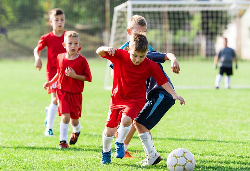
- Adolescents taking part in sports that involve lots of running and jumping are thought to be most affected.
- Localised pain on the front of the shin, below the kneecap.
- Pain that is aggravated by activity, specifically activities such as sprinting, running and jumping (3).
- A palpable lump below the kneecap, on the top and centre of the shin bone. This is also usually the site of pain.
- If you are concerned, a musculoskeletal physiotherapist can provide a diagnosis and advise on how to manage symptoms.
- Reduce or modify levels of activity.
- In severe cases, ceasing activity for a period of time might be required.
- Simple exercises to help improve the strength and flexibility of muscles around the knee joint.
- Overall, the condition has an excellent long-term prognosis.
- The condition can take up to 2 years to fully settle, but often resolves itself within a matter of weeks or months.
- 90% of patients who experience the condition make a full recovery as they finish their active growth spurt (3,4).
We recommend consulting a musculoskeletal physiotherapist to ensure exercises are best suited to your recovery. If you are carrying out an exercise regime without consulting a healthcare professional, you do so at your own risk. Book online with us today to get a programme tailored to your specific needs.
2. Signs and Symptoms
- Localised pain on the tibial tuberosity, below the patella (3).
- Swelling and a bony lump around the tibial tuberosity (3).
- Symptoms are commonly aggravated by activities such as weight training, sprinting/running and jumping.
- Symptoms are often eased by rest.
3. Causes
Osgood-Schlatter disease can be defined as an irritation caused by traction (a pulling force) to the top of the tibia (shin bone) and irritation of the patella tendon. During periods of rapid growth, our bones often grow at a quicker rate than our muscles, so when our bones grow our muscles have to temporarily stretch to account for this increased length of the bone. In patients with Osgood-Schlatter disease, this excess load applies traction to the tibial tuberosity, which lies directly over a growth plate in growing bones. This, combined with increases in load through the area such as when running and jumping, can result in irritation of the attachment site, tendon and growth plates (2,3).
4. Risk Factors
This is not an exhaustive list. These factors could increase the likelihood of someone developing Osgood-Schlatter disease. It does not mean everyone with these risk factors will develop symptoms.
- Young age – seen more frequently in active children and adolescents, particularly in boys aged 12 to 15-years-old, and girls aged 8 to 12-years-old (3).
- Sports – more common in sports which include lots of running, jumping or landing, such as football, basketball, netball or athletics.

5. Prevalence
Most commonly occurs in adolescents. 10% of adolescents are affected and the prevalence is more common in adolescents that are active (4).
6. Assessment & Diagnosis
Musculoskeletal physiotherapists and other appropriately qualified healthcare professionals can provide you with a diagnosis by obtaining a detailed history of your symptoms. A series of physical tests might be performed as part of your assessment to rule out other potentially involved structures and gain a greater understanding of your physical abilities to help facilitate an accurate working diagnosis.
Your treating clinician will want to know how your condition affects you day-to-day so that treatment can be tailored to your needs and personalised goals can be established. Intermittent reassessment will ascertain if you are making progress towards your goals and will allow appropriate adjustments to your treatment to be made. Imaging studies such as MRIs, X-rays and ultrasounds are rarely needed in the diagnosis but in unusual presentations, they may be called for.
7. Self-Management
As Osgood–Schlatter disease is a self-limiting condition, self-management plays a key role in the management of symptoms. As part of your treatment and rehabilitation, a musculoskeletal physiotherapist or other appropriately qualified healthcare professional will help you understand your condition and what you can do to effectively manage symptoms.
8. Rehabilitation
The recovery from this condition revolves mainly around reducing the stress to allow the tissues to settle down or the rate of growth to reduce. However, rehabilitation is important particularly to reduce any deconditioning during the recovery process.
9. Osgood-Schlatter Disease
Rehabilitation Plans
Our team of expert musculoskeletal physiotherapist have created rehabilitation plans to enable people to manage their condition. If you have any questions or concerns about a condition, we recommend you book an consultation with one of our clinicians.
What Is the Pain Scale?
The pain scale or what some physios would call the Visual Analogue Scale (VAS), is a scale that is used to try and understand the level of pain that someone is in. The scale is intended as something that you would rate yourself on a scale of 0-10 with 0 = no pain, 10 = worst pain imaginable. You can learn more about what is pain and the pain scale here.
90% of cases will resolve independently of treatment. However, low-intensity loading/flexibility work is known to improve pain (3, 6). Combining light stretching exercises with pain management strategies helps offload the aggravated tendon, whilst reducing pain. Pain should not exceed 3/10 on your self-perceived pain scale whilst completing this exercise programme.
- 0
- 1
- 2
- 3
- 4
- 5
- 6
- 7
- 8
- 910
Once symptoms begin to settle, you can gradually increase the levels of activity participation within the limits of your pain. This may include a gradual increase in training capacity (going from 2 to 4 days a week) and/or a gradual reintroduction of lower limb weight/resistance training with low weight/resistance. You may also be able to start taking part in light training drills. Discussions with coaches about how to integrate training is recommended. Avoiding the ‘boom and bust’ cycle by avoiding a rapid increase in training load or activity and gradually increasing your activity allows your muscles and tendons to adapt to the new level of activity. Rapidly increasing training frequency or intensity can result in overloading the muscles and tendons which can cause your symptoms to flare up. It is also important to allow for adequate rest between training/activity days to allow for the growth and repair of muscles and tendons after activity. Pain should not exceed 4/10 on your self-perceived pain scale whilst completing this exercise programme.
- 0
- 1
- 2
- 3
- 4
- 5
- 6
- 7
- 8
- 910
At the end stage of rehabilitation, near full levels of activity can be resumed within the constraints of your symptoms. A gradual increase in your levels of regular activity can be completed, as well as an increase in the intensity of your training or activity. More intense strength work can be included into your rehabilitation programme once pain has settled and you have made a successful return to sport. Pain should not exceed 4/10 on your self-perceived pain scale whilst completing this exercise programme.
- 0
- 1
- 2
- 3
- 4
- 5
- 6
- 7
- 8
- 910
10. Return to Sport / Normal life
For patients wanting to achieve a high level of function or return to sport we would encourage a consultation with a physiotherapist as you will likely require further progression beyond the advanced rehabilitation stage.
As part of a comprehensive treatment approach, your musculoskeletal physiotherapist may also use a variety of other pain relieving treatments to support symptom relief and recovery. Whilst recovering you might benefit from a further assessment to ensure you are making progress and establish the appropriate progression of treatment. Ongoing support and advice will allow you to self-manage and prevent future re-occurrence.
11. Other Treatment Options
- Surgery is very rarely needed and would usually not be carried out until the child’s growth plates have closed.
- Soft tissue treatments such as sports massage may provide temporary pain relief but should not be used as a stand-alone treatment (1).
12. Links for Further Reading
25 locations and counting across the UK
References
- Crawford, C. et al. (2016). ‘The Impact of Massage Therapy on Function in Pain Populations-A Systematic Review and Meta-Analysis of Randomized Controlled Trials: Part I, Patients Experiencing Pain in the General Population.’, Pain medicine (Malden, Mass.), doi: 10.1093/pm/pnw099. 17, 1353–1375.
- Dobbe, A. M. and Gibbons, P. J. (2017). ‘Common paediatric conditions of the lower limb’, Journal of Paediatrics and Child Health, doi: 10.1111/jpc.13756. 53, 1077–1085.
- Gholve, P. A. et al. (2007). ‘Osgood Schlatter syndrome.’, Current opinion in pediatrics, doi: 10.1097/MOP.0b013e328013dbea. 19, 44–50.
- De Lucena, G. L., dos Santos Gomes, C. and Guerra, R. O. (2011). ‘Prevalence and associated factors of Osgood-Schlatter syndrome in a population-based sample of Brazilian adolescents.’, The American journal of sports medicine, doi: 10.1177/0363546510383835. 39, 415–420.
- Magrini, D. and Dahab, K. S. (2016). ‘Musculoskeletal Overuse Injuries in the Pediatric Population’, Current Sports Medicine Reports, Available at: https://journals.lww.com/acsm-csmr/Fulltext/2016/11000/Musculoskeletal_Overuse_Injuries_in_the_Pediatric.8.aspx. 15.
- Vaishya, R. et al. (2016). ‘Apophysitis of the Tibial Tuberosity (Osgood-Schlatter Disease): A Review’, Cureus, 8.
Other Conditions in
Knees, Paediatrics
Patellofemoral Pain Syndrome (PFPS)
Knee pain around the kneecap usually worse in static positions, squatting or kneeling.
Patellar Tendinopathy
Knee pain at the lower border of the kneecap which is also known as ‘jumper’s knee’.
Patella Dislocation
Patella dislocation is a knee injury in which the patella (kneecap) slips out of its normal position. The most common direction for the kneecap to dislocate is laterally or the outside. This is commonly associated with pain and swelling in the soft tissue tissues which may have been stretched or damaged. Patella subluxation refers to when the kneecap is only partially displaced and then returns to it’s normal location.
Meniscus Injury
Structural knee injury, triggered either by a tear or through wear and tear.
Medial Collateral Ligament Sprain
The medial collateral ligament is on the inner side of the knee. It provides stability to the joint by preventing excessive side–to–side movement. It is possible to injure this ligament when a person is bearing weight, and the knee is forced inwards.
Lateral Collateral Ligament (LCL) Injury
The lateral collateral ligament is a strong ligament on the outside of the knee. A tear will only occur during a high force impact or twisting motion.
Knee Replacement Surgery
Replacement of the knee hinge joint, typically as a result of severe osteoarthritis or trauma.
Knee Osteoarthritis
Common age related changes to the structure of the knee joint which may be associated with pain, stiffness and loss of function.
Iliotibial Band Syndrome
Presents as pain on the outside of the knee, normally occurring because of overload due to prolonged or repeated bouts of exercise.
Hamstring Strain/Tear
An over-stretch or tear to one or more of the muscles located at the back of the thigh.
Femoral Nerve Radiculopathy
This is where the nerve that supplies the front of the leg is irritated and causes pain/numbess.
Fat Pad Impingement
A rare condition affecting the adipose (fat) tissue that sits under the kneecap (patella) between the joint spaces of the knee.
Degenerative Meniscus
Seen to be normal as we age, but in some situations can result in knee aches, pain or joint swelling.
Bowed Knees
A condition in which the legs are bowed outwards leaving a greater space in between your knees.
Benign Joint Hypermobility Syndrome
Common age related changes to the structure of the knee joint which may be associated with pain, stiffness and loss of function.
Baker’s Cyst
Swelling in the popliteal space (space behind the knee) that causes a visible lump.
Anterior Cruciate Ligament (ACL) Injury
Injury to a major stability ligmant in the knee, normally occuring following a significant twisting injury.