Hip Osteoarthritis
1. Introduction
Osteoarthritis occurs when the smooth protective cartilage lining of a joint becomes irritated or thins naturally over time. This is a predictable and normal part of the ageing process and does not mean that we will all suffer from joint pain or osteoarthritis. Osteoarthritis is the medical term to describe when age-related changes to our joints result in pain, restricted motion and functional difficulties (2, 3). It is a common and disabling condition, resulting in significant medical costs and adversely affecting the person’s quality of life. However, research continues to demonstrate the most effective ways of managing osteoarthritis. As a result, there are several treatment approaches that can help reduce the impact of the condition on daily life.
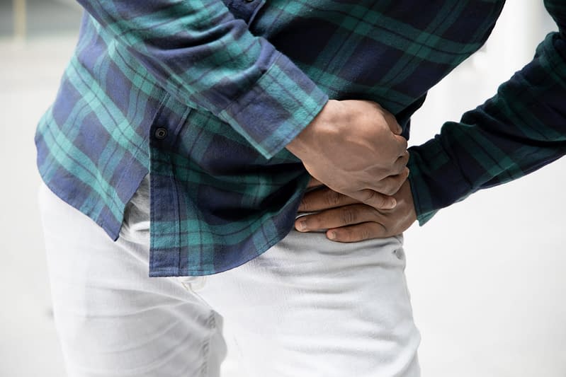
Osteoarthritis of the hip refers to changes in the structure and function of the hip joint, resulting in hip/groin pain, reduced range of movement and muscle weakness. This can result in mild symptoms, to more severe forms of the disease. It is worth noting that the degree of change on X-ray often relates quite poorly to the level of pain a person is experiencing. It is also worth noting that for most people, their symptoms and progression of joint change remain similar for many years.
Frequently Asked Questions
- Hip joint osteoarthritis involves changes to the structure of the hip joint and may be associated with pain, stiffness and loss of function of the joint.
- Common – most adults have signs of age-related changes to the hip joint on X-ray, yet many will have no pain or stiffness of the joint (asymptomatic).
- 10% of adults over the age of 20 in the UK are affected by symptomatic osteoarthritis in at least one joint (1).
- The hip is the second most common site of symptomatic osteoarthritis after the knee (1).
- No.
- Hip joint osteoarthritis is not directly linked to any other serious pathology.
- With the correct assessment and management, osteoarthritic joints can be well managed (2, 4, 5).
- Typically, adults over 45 years of age are most likely to be affected.
- Increasing rates of hip joint osteoarthritis are seen with increasing age (2).
- Younger patients with a history of childhood hip trauma or injury may develop earlier onset osteoarthritis.
- Hip joint osteoarthritis is slightly more common in women (12.8%) than men (8.6%) (1).
- Pain and stiffness in the hip or groin, particularly in the mornings or after prolonged periods of being stationary.
- Groin pain and difficulty crossing legs or putting on shoes and socks.
- Stiffness and popping sounds/feelings when moving the joint.
- Fatigue, as pain experienced day-to-day can be tiring.
- Maintain a healthy weight and body mass index (BMI).
- Exercise, particularly aerobic exercise and exercises to strengthen the hip joint.
- Pain and anti-inflammatory medication may help relieve pain in the short term.
- Corticosteroid injections may be indicated for some people.
- Joint replacement surgery may be offered to those in severe pain, or where all other treatment has failed.
- Although there is no “cure” for osteoarthritis, sensible management can significantly improve the symptoms associated with it.
- Regular exercise, weight management and a healthy lifestyle will minimise the impact of hip osteoarthritis.
We recommend consulting a musculoskeletal physiotherapist to ensure exercises are best suited to your recovery. If you are carrying out an exercise regime without consulting a healthcare professional, you do so at your own risk. Book online with us today to get a programme tailored to your specific needs.
2. Signs and Symptoms
- Pain in the hip, groin or down the front of the thigh to the knee.
- Hip joint stiffness – particularly in the morning or after prolonged sitting.
- Crepitus- crackling or popping sounds when moving the affected joint.
- It is worth repeating that many people have X-ray changes that indicate some degree of osteoarthritis but have no, or only very mild, symptoms from the above.
3. Causes
How hip joint osteoarthritis develops into a painful disorder for some people and not others is not fully understood but it certainly seems that it is not purely a case of gradual “wear and tear”. All normal joints and tissues are constantly undergoing some form of repair or regeneration because of the normal stress and strain of daily life. It seems that osteoarthritis develops when the body’s repair or regenerative processes cannot keep up with the changes over time due to the ageing process.
4. Risk Factors
This is not an exhaustive list. These factors could increase the likelihood of someone developing symptomatic hip joint osteoarthritis. It does not mean everyone with these risk factors will develop symptoms.
- Age – osteoarthritis becomes more common with increasing age due to the natural regenerative mechanisms becoming less efficient in some people as they age.
- Family history – there may be some inherited tendency for osteoarthritis to develop in some people if relatives or parents have had a similar disease.
- Obesity – osteoarthritis is more likely to develop, or be more severe, in obese people due to increased load on the joints and the potential for more pronounced joint changes.
- Gender – women (12.8%) are more likely to develop osteoarthritis than men (8.6%) (1).
- Previous joint injury, damage, or deformity – for example, joint infection, a previous break (fracture) in the bone around a joint, or a previous ligament injury that caused joint instability.

5. Prevalence
In the UK, approximately 10% of adults have symptomatic, clinically diagnosed osteoarthritis, the knee being the most common, followed closely by the hip (1). Changes in imaging of the hip joint are very common and most adults (over 35) have signs of changes on X-ray. However, most people with X-ray findings of osteoarthritis do not have symptoms and indeed, may never develop symptoms. There is a very poor correlation between what we see on an X-ray or magnetic resonance image (MRI) scan and a person’s symptoms. This means that even people with severe radiographic (appearance of the hip on an X-ray) changes may be relatively unaffected by pain or joint stiffness throughout their life.
It is important not to worry about being told you have osteoarthritis as it is very common, and we now know that it often does not cause increasing pain, stiffness or disability. There is good research showing that sensible management can have a significant impact on reducing the effects of osteoarthritic hip pain on your life. (2, 3, 5, 6).
6. Assessment & Diagnosis
It is important to get an accurate diagnosis if you think you have osteoarthritis, as there are different types of arthritis that may need different treatments (3). The diagnosis of osteoarthritis is usually based on the physiotherapist taking a detailed history of your symptoms, including how and when they started, how they have developed, how they affect your life and any factors that make them better or worse. This is usually followed by a physical examination where your doctor or physiotherapist will assess for tenderness around the hip joint, examine the movements of the joint which may be sore and stiff, and look for any weakness of the muscles that support the joint.
Other tests such as X-ray imaging or blood tests may help in the diagnosis of osteoarthritis but are often not required. They may, however, be used in some cases where the diagnosis is uncertain, or to rule out other issues.
7. Self-Management
Although there is no cure for osteoarthritis, there is a range of strategies that can help you to manage pain and optimise your function (2, 3, 4, 5, 6). These include:
- Regular and appropriate exercise – aerobic exercise such as walking, cycling or swimming may be the best for pain and function improvements (2, 3, 5, 6).
- Strengthening the muscles around the hip, pelvis and lower back. Strength and stability can have a very positive impact on symptoms (2, 4, 6).
- Maintaining a healthy body weight to reduce stress on the hip joints (2, 3, 4, 6).
- Pain relief medications – paracetamol, ibuprofen gel (in agreement with your GP).
- Heat or ice treatment – applying a hot-water bottle or ice (wrapped in a towel to protect your skin) may help to ease the pain. Do this for 20 mins, allowing for 2 hours off (3).
- Footwear – in general, the ideal shoe would have a thick but soft sole, soft uppers and plenty of room at the toes and the ball of the foot. (3, 6).
- Pacing yourself – if there are jobs that often increase your pain, try to break them down and allow time for rest breaks (3, 6).
8. Rehabilitation
Although it may not be possible to stop osteoarthritic changes from occurring, there is scope to limit the progression with appropriate management. Guided rehabilitation should look to incorporate several strategies such as activity modification, self-awareness and exercise therapy with the goal of helping you to manage your issue, limit further negative joint changes and improve your function. For maximal impact, such strategies should be combined with manual techniques that can be performed by your physiotherapist to help improve the function of your hip (7, 8).
Osteoarthritis is more common as you get older, but it is not just part of getting older. You do not have to live with pain or disability. Various levels of rehabilitation exercises should be considered based on your ability.
9. Hip Osteoarthritis
Rehabilitation Plans
Our team of expert musculoskeletal physiotherapist have created rehabilitation plans to enable people to manage their condition. If you have any questions or concerns about a condition, we recommend you book an consultation with one of our clinicians.
What Is the Pain Scale?
The pain scale or what some physios would call the Visual Analogue Scale (VAS), is a scale that is used to try and understand the level of pain that someone is in. The scale is intended as something that you would rate yourself on a scale of 0-10 with 0 = no pain, 10 = worst pain imaginable. You can learn more about what is pain and the pain scale here.
Exercise rehabilitation for hip osteoarthritis involves a progression from a gentle range of motion exercises to help restore and maintain the movement of your hip, to strength-based exercises to ensure the surrounding muscles are strong enough to adequately support and protect the joint (7, 8). In the earlier stages of treatment, stretching exercises that target tighter muscles around the hip joint have been shown to improve function and reduce pain (8). Pain should not exceed 4/10 on your perceived pain scale whilst completing this exercise programme.
- 0
- 1
- 2
- 3
- 4
- 5
- 6
- 7
- 8
- 910
In the intermediate stages, rehabilitation should look to shift from predominantly stretching exercises to incorporate more strength-orientated exercises. Commonly these begin with more simple exercises involving the muscles that support the hip, before aiming to become more functional (relating to activities of daily living) in the later stages (7, 8). Pain should not exceed 4/10 on your perceived pain scale whilst completing this exercise programme.
- 0
- 1
- 2
- 3
- 4
- 5
- 6
- 7
- 8
- 910
By this point, you will have worked on restoring the range of motion in the affected hip and strengthening the supporting muscles. This is the time to make the programme more specific to activities of daily living such as walking, climbing stairs and bending amongst others. Pain should not exceed 4/10 on your perceived pain scale whilst completing this exercise programme.
- 0
- 1
- 2
- 3
- 4
- 5
- 6
- 7
- 8
- 910
10. Return to Sport / Normal life
For patients wanting to achieve a high level of function or sport, we would encourage a consultation with a physiotherapist as you will likely require further progression beyond the advanced rehabilitation stage.
As part of a comprehensive treatment approach, your musculoskeletal physiotherapist may also use a variety of other pain-relieving treatments to support symptom relief and recovery. Whilst recovering you might benefit from a further assessment to ensure you are making progress and establish the appropriate progression of treatment. Ongoing support and advice will allow you to self-manage your condition.
11. Other Treatment Options
In some cases, the management strategies outlined above may not be enough to help people manage their problems from day-to-day. In these cases, options for escalated care do exist. These may include a trial of corticosteroid into the joint. Whilst careful consideration should be made regarding possible side effects or long-term reliance on corticosteroid injections, they can be a useful tool to control pain, allowing for the commencement of exercise and lifestyle rehabilitation.
If corticosteroid injections fail to facilitate a return to adequate function, your GP or specialist physiotherapist may look to refer you to an orthopaedic consultant for consideration of joint replacement surgery if you fit the referral criteria.
25 locations and counting across the UK
References
- Swain, S., Sarmanova, A., Mallen, C., Kuo, C.F., Coupland, C., Doherty, M. and Zhang, W., 2020. Trends in incidence and prevalence of osteoarthritis in the United Kingdom: findings from the Clinical Practice Research Datalink (CPRD). Osteoarthritis and cartilage, 28, 792-801.
- The National Health Service. 2018. Arthritis: Overview. Available at: https://www.nhs.uk/conditions/arthritis/.
- Versus Arthritis. (2020) Osteoarthritis of the hip information booklet. Available at: https://www.versusarthritis.org/media/22728/osteoarthritis-of-the-hip-information-booklet.pdf
- Loeser, R.F., Collins, J.A. and Diekman, B.O. (2016) Ageing and the pathogenesis of osteoarthritis. Nature Reviews Rheumatology, 12, 412-420.
- Goh, S.L., Persson, M.S., Stocks, J., Hou, Y., Welton, N.J., Lin, J., Hall, M.C., Doherty, M. and Zhang, W., 2019. Relative efficacy of different exercises for pain, function, performance and quality of life in knee and hip osteoarthritis: systematic review and network meta-analysis. Sports Medicine, 49, 743-761.
- Bennell, K. (2013). Physiotherapy management of hip osteoarthritis. Journal of physiotherapy, 59, 145-157.
- Hurley, M., Dickson, K., Hallett, R., Grant, R., Hauari, H., Walsh, N., Stansfield, C. and Oliver, S. (2018). Exercise interventions and patient beliefs for people with hip, knee or hip and knee osteoarthritis: a mixed methods review. Cochrane Database of Systematic Reviews.
- Bennell, K. (2013) Physiotherapy management of hip osteoarthritis. Journal of physiotherapy, 59, 145-157.
Other Conditions in
Hips & Pelvis, Long Term Conditions, Orthopaedics, Pain
Sacroiliac Joint Dysfunction
Pain originating from the sacroiliac joint at the base of your back where the spine joins the pelvis.
Proximal Hamstring Tendinopathy
Pain and weakness under the buttock or the back of your upper thigh caused by tendon issues.
Pelvic Girdle Pain
Typically seen in pregnancy causing pain, instability and limitation of mobility and functioning of the pelvic joints.
Pelvic Floor Dysfunction
The inability to effectively control the muscles of your pelvic floor, leading to issues with continence and pain.
Meralgia Paraesthetica
Pain on the outside thigh caused by compression and inflammation of the nerve that supplies that area
Lumbar Disc Injury
Lumbar discs sit between each of the bones of the spine. Problems can occur when these discs become irritated.
Low Back Pain and Sciatica
Sciatica is a symptom describing pain and/or pins and needles down the back of the leg.
Iliotibial Band Syndrome
Presents as pain on the outside of the knee, normally occurring because of overload due to prolonged or repeated bouts of exercise.
Hip Replacement Surgery
Replacement of the hip ball and socket joint, typically as a result of severe osteoarthritis or trauma.
Hamstring Strain/Tear
An over-stretch or tear to one or more of the muscles located at the back of the thigh.
Greater Trochanteric Pain Syndrome
A condition affecting the tendons that insert into outside of the hip. A common cause of pain felt around the hip and pelvis.
Femoroacetabular Impingement
A condition that results in pain in the groin, hip and down the front of the thigh.
Femoral Nerve Radiculopathy
This is where the nerve that supplies the front of the leg is irritated and causes pain/numbess.
Coccydynia
Coccydynia is the medical term used to describe pain in your coccyx (tail bone).
Benign Joint Hypermobility Syndrome
Common age related changes to the structure of the knee joint which may be associated with pain, stiffness and loss of function.
Adolescent Hip Dysplasia
A result of an abnormality of the hip joint anatomy resulting in pain in the hip with occasional instability.
Adductor-Related Groin Pain
Localised discomfort to the inner upper thigh and groin.